RNA Fragments Protect Neurons from Botulinum Toxin by Blocking Cell Death Pathway
Israeli and Hebrew University researchers discover molecular mechanism that keeps nerve cells alive during botulinum poisoning, potentially extending therapeutic applications
In a discovery that could revolutionize both medical treatments and cosmetic procedures, scientists have identified how neurons survive exposure to botulinum toxin—one of the most potent biological toxins known to humans. Researchers from the Israel Institute for Biological Research and Hebrew University of Jerusalem found that poisoned neurons produce specific RNA fragments that block a cell death pathway called ferroptosis, keeping the cells alive despite paralysis. The findings, published today in Genomic Psychiatry, explain a decades-old mystery about why botulinum toxin causes temporary paralysis without killing nerve cells.
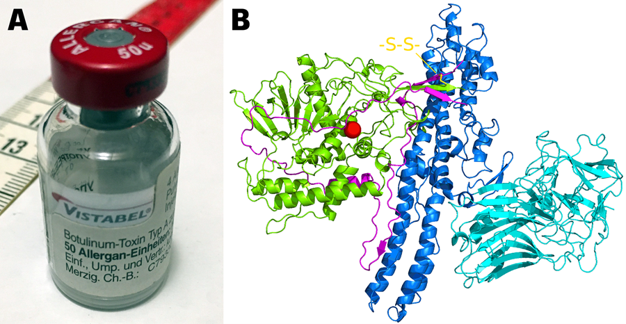
Figure 1. (A) Medical vial containing botulinum toxin Type A (Vistabel) for cosmetic use, shown with centimeter ruler for scale. Credit: Pittigrilli, CC BY 4.0. (B) Crystal structure of botulinum toxin A1 showing light chain (green) with zinc cofactor (red), heavy chain translocation domain (blue), and receptor-binding domain (cyan), connected via disulfide bond (yellow). PDB: 3BTA. Credit: 5-HT2AR, CC0 Public Domain.
The Study Design
The research team, led by Arik Monash and colleagues, exposed human neuroblastoma cells to lethal doses of botulinum neurotoxin A (BoNT/A)—approximately 300 times the concentration used in cosmetic Botox treatments. They studied these cells over 48 hours, tracking molecular changes using advanced RNA sequencing technology.
The study involved comparing poisoned neurons to healthy control cells, analyzing over 12 million RNA sequences per sample. Researchers also validated their findings using rat submandibular glands exposed to the toxin, confirming the response occurs across different mammalian tissues.
"We wanted to understand not just how the toxin blocks nerve signals, but why neurons don't die from this extreme stress," explained senior author Professor Hermona Soreq. "What we found was an elegant survival mechanism that cells activate specifically under botulinum exposure."
What They Found
The key discoveries include:
• 335 different transfer RNA fragments (tRFs) changed dramatically in poisoned neurons, with 63% increasing in abundance • The most elevated fragment, 5'LysTTT, increased over 4000% compared to healthy cells
• 20% of elevated tRFs contained an identical 11-letter genetic code that appears to coordinate the survival response • Ferroptosis markers decreased by 50% despite conditions that normally trigger this cell death pathway
These RNA fragments work like molecular switches, turning off death signals while maintaining basic cellular functions. Unlike the minor changes seen in microRNAs—previously thought to be the main RNA regulators—these tRNA fragments showed massive alterations specifically triggered by botulinum exposure.
Why It Works
Think of ferroptosis like rust forming on iron—cells accumulate damaging oxidized fats until they essentially corrode and die. Normally, when neurons are stressed by toxins, this rusting process accelerates rapidly.
The botulinum-triggered RNA fragments act like a protective coating, specifically the 5'LysTTT fragment. This fragment binds to and silences CHAC1, a protein that normally promotes ferroptosis. With CHAC1 suppressed, cells maintain their antioxidant defenses despite the toxin's presence.
What makes this discovery unique is the coordinated response: hundreds of RNA fragments working together, many sharing the same regulatory sequence. This amplifies their protective effect far beyond what individual molecules could achieve.
What This Means for Patients
Currently, patients receiving botulinum treatments for conditions like dystonia, chronic migraines, or excessive sweating require injections every 3-4 months. The temporary nature of the treatment—while frustrating for some patients—actually depends on neurons surviving to eventually recover function.
Understanding this survival mechanism could enable development of longer-lasting formulations. For someone with cervical dystonia who currently needs quarterly injections, extended treatments could mean fewer medical visits, reduced healthcare costs, and improved quality of life.
The timeline for clinical applications could be 5-10 years, pending further research and regulatory approval. Initial applications would likely focus on extending the duration of existing treatments rather than developing entirely new therapies.
From Lab to Clinic
The next steps involve testing whether manipulating these RNA fragments can control treatment duration. Researchers plan to investigate whether enhancing tRF production could extend therapeutic effects to 6 months or longer.
The regulatory pathway would require demonstrating safety in animal models before human trials. Since botulinum treatments are already FDA-approved, modifications to extend duration might follow an accelerated approval process.
Cost considerations remain important—longer-lasting treatments could reduce per-year expenses despite potentially higher per-dose costs. The technology could particularly benefit patients in remote areas with limited access to specialty clinics.
Bigger Picture Impact
From a healthcare system perspective, extending botulinum treatment duration could save millions in medical visits and lost productivity. A patient receiving treatment for spasticity might reduce clinic visits from four to two annually, freeing healthcare resources for other needs.
Scientifically, this discovery opens new research directions in neurodegeneration and cell death. The same protective mechanisms might apply to conditions like Parkinson's disease or ALS, where ferroptosis contributes to neuron loss. The identified RNA fragments could serve as templates for developing neuroprotective drugs beyond botulinum applications.
Important Caveats
Researchers note several limitations to consider. The study used extremely high toxin concentrations that don't reflect cosmetic uses, where doses are roughly 300 times lower. The protective mechanisms identified may not activate at these minimal doses.
The research examined cells for only 48 hours, while clinical effects last months. Longer-term studies are needed to confirm these mechanisms persist throughout the treatment duration. Additionally, the cell line used, while human-derived, may not perfectly represent mature neurons at neuromuscular junctions.
Independent Perspectives
While this study presents compelling findings, independent validation from other research groups will be essential. External perspectives from neurotoxin researchers, clinical neurologists, and patient advocacy groups would provide valuable context about the potential clinical implications and necessary safety considerations for any future therapeutic applications based on these mechanisms.
The findings warrant replication in different cell types and animal models before conclusions can be drawn about human therapeutic potential.
Next Steps
The immediate research priorities include:
• Testing tRF responses in human motor neurons derived from stem cells • Investigating whether tRF levels correlate with treatment duration in patients
• Developing methods to enhance or suppress specific tRFs • Exploring applications for other neuromuscular conditions • Examining whether similar mechanisms exist for other BoNT serotypes
The Bottom Line
This research reveals an elegant molecular dance where poisoned neurons activate hundreds of protective RNA fragments to survive one of nature's deadliest toxins. Rather than simply enduring the assault, cells mount a sophisticated defense that blocks multiple death pathways while maintaining essential functions. By understanding this natural survival program, scientists now have a roadmap for potentially extending botulinum treatments from months to half a year or longer—a development that could benefit millions receiving these therapies worldwide. The next milestone will be demonstrating that manipulating these RNA fragments in living organisms can safely extend treatment benefits without compromising the precision that makes botulinum therapies so valuable.
Content Details
The peer-reviewed article appears in Genomic Psychiatry, providing valuable context about Genomic Press's mission to support innovative, cross-disciplinary research bridging fundamental neuroscience and translational initiatives in brain medicine.
🔓🆓 Open Access • Genomic Psychiatry
Peer-reviewed Research Article: https://doi.org/10.61373/gp025a.0047
Corresponding Authors: Joseph Tam (yossi.tam@mail.huji.ac.il), Osnat Rosen (osnatr@iibr.gov.il), Hermona Soreq (hermona.soreq@mail.huji.ac.il)
Funding: Israel Science Foundation Grants 835/23 & 1266/24
Conflicts of Interest: None declared
📱 Social Media Links:
✖️ X (Twitter): https://x.com/GenomicPress/status/1973833813869359499
🔗 LinkedIn: https://lnkd.in/dwr4pJGp
📸 Instagram: https://www.instagram.com/p/DPUbc4zCSPk/
📘 Facebook: https://www.facebook.com/share/p/174wu77kVP/
🦋 Bluesky: https://bsky.app/profile/genomicpress.bsky.social/post/3m2a7i7qjvs2a
🌎 External Coverage
🇺🇸 EurekAlert! press release: https://www.eurekalert.org/news-releases/1084279
About This Publication
This groundbreaking study exemplifies the world-class research published in Genomic Psychiatry, where innovative molecular investigations are reshaping our understanding of neurotoxin biology and therapeutic applications. By providing an open-access platform for transformative neuroscience research, Genomic Psychiatry continues to establish itself as the definitive venue for innovative neuroscience.